Skip to content

Explore Our Research

Decoding PNPLA3-Mediated Pathogenesis in MASLD

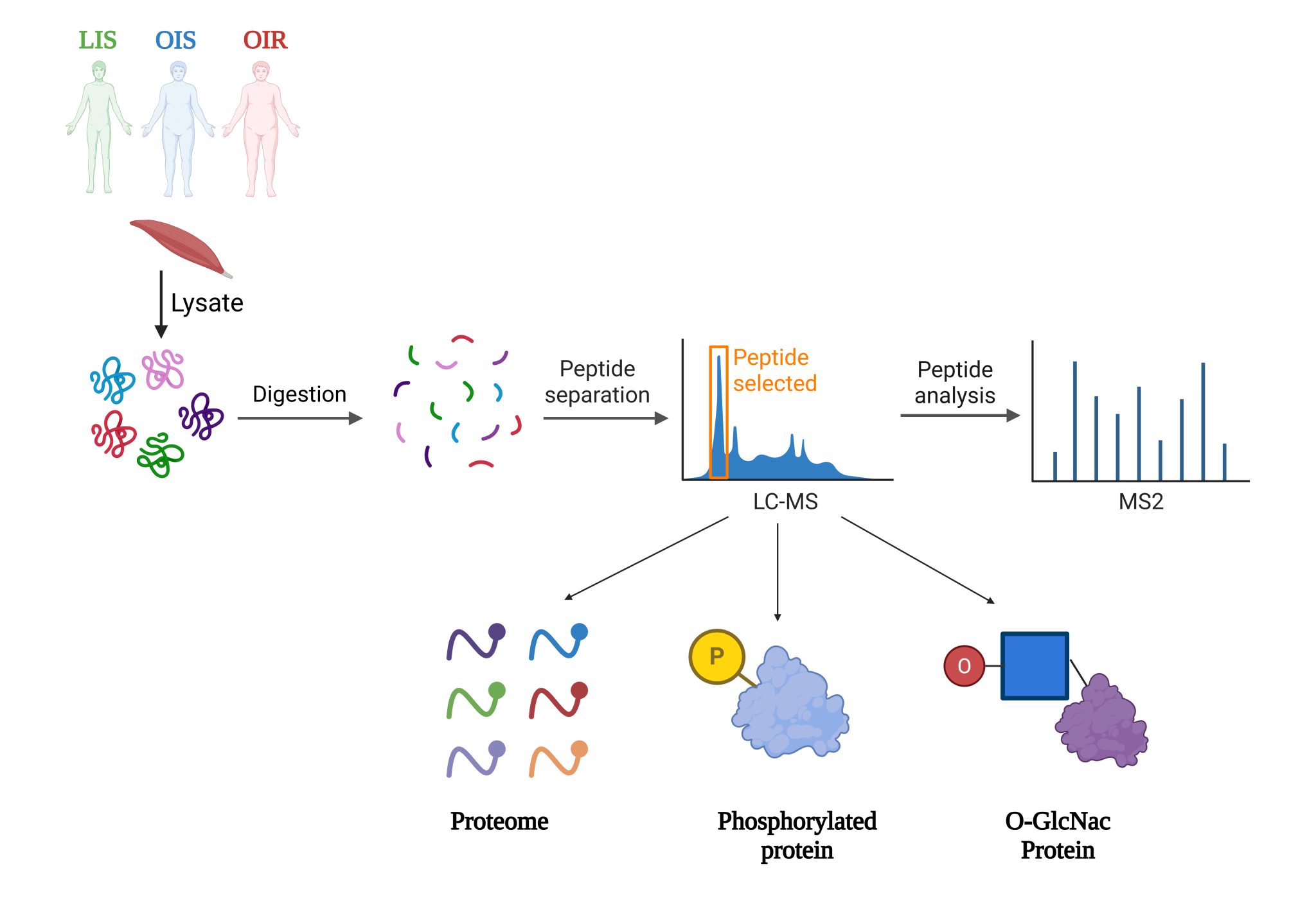
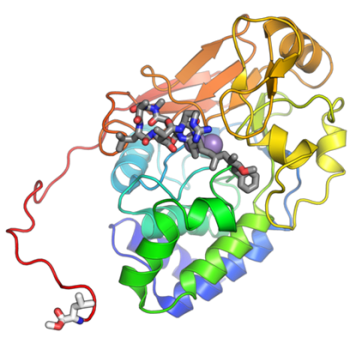
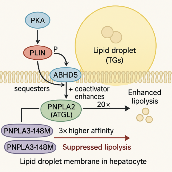
Our PNPLA3 project investigates the molecular mechanisms by which the I148M mutation in PNPLA3 contributes to the development of Metabolic Associated Steatotic Liver Disease (MASLD). Using integrative approaches, including protein-protein docking, molecular dynamics (MD) simulations, and LC-MS/MS-based proteomics & interactomics, we demonstrate that PNPLA3-I148M competitively sequesters the coactivator ABHD5 away from its primary lipase partner, PNPLA2 (ATGL). This impaired lipolytic signaling leads to hepatic triglyceride accumulation—a key hallmark of MAFLD. Ongoing efforts are focused on identifying potential small-molecule modulators of PNPLA3-ABHD5 interactions to restore lipolysis and provide novel therapeutic strategies for fatty liver disease.

Skeletal Muscle Modifications in Obesity and Insulin Resistance

This study investigates how proteins and their modifications—Phosphorylation and O-glycosylation—differ in skeletal muscle between lean and obese people with variable insulin sensitivity. By studying these molecular patterns, this work offers promising results to uncover pathways that contribute to insulin resistance and potential protective factors in obesity. 

Studying how the development of insulin resistance is impacted by Protein Phosphatase 2A

This study is observing how protein phosphatase 2A (PP2A) affects the body during the development of insulin. PP2A is a highly regulated phosphatase that plays a role in the insulin stimulated pathway through the removal of phosphates from key proteins invloved within the pathway. This research will be done by studying PP2A and its interaction partners as well as its post-translational modifications using human skeletal muscle tissue samples and analyzing the data with our HPLC-ESI-MS/MS. Overall, this study will give us a better understanding of the development of insulin resistance and the role of PP2A during this development.

Proteomic Discovery of Bifunctional Apoptosis Regulator Substrates

We will compare BFAR-dependent ubiquitination sites between wild-type and BFAR knockdown Huh7 cells using anti-diGly (K-8-GG) enrichment. We will perform ubiquitin remnant (K-ε-GG) enrichment using the Cell Signaling Technology PTMScan® Ubiquitin Enrichment Kit and multiplexed quantification using TMT labeling. We will determine how BFAR loss affects global protein expression and pathways using Tandem Mass Tag (TMT). To simulate metabolic stress and facilitate the identification of lipid-sensitive BFAR substrates, cells will be treated with lipids. This integrative strategy broadens our understanding of BFAR’s role in lipid metabolism and proteostasis by allowing us to investigate both BFAR-dependent ubiquitination events and global proteome alterations.